Loading...
About services
Our services
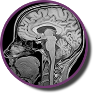
Head MRI
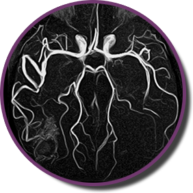
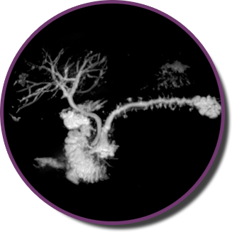
Head MRI + angiography
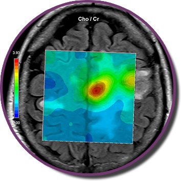
Head MRI with spectroscopy
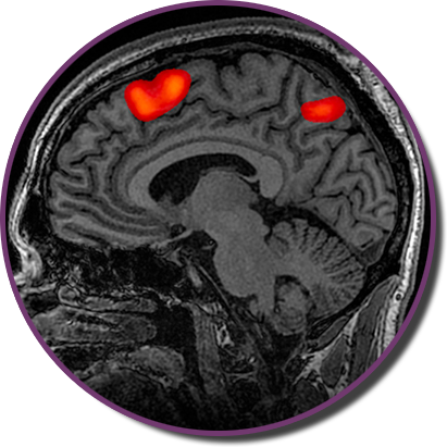
Functional MRI
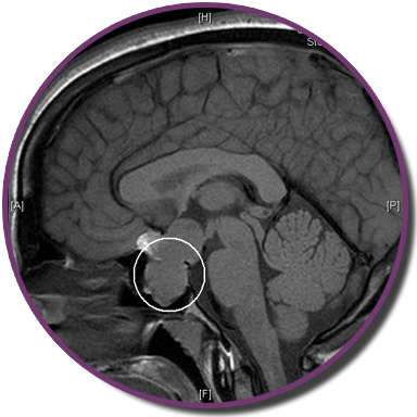
Hypophysis MRI
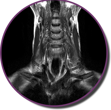
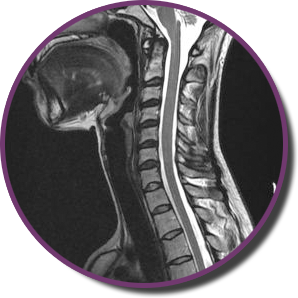
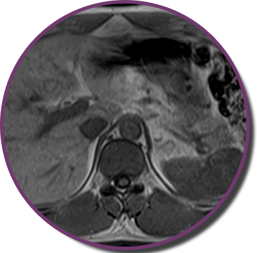
Neck MRI
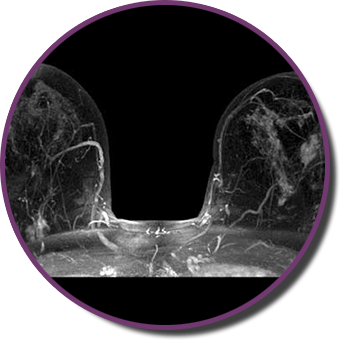
Breast MRI
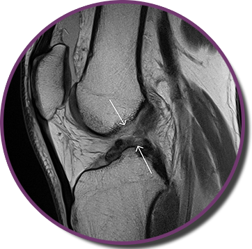
Musculoskeletal MRI
Spine MRI
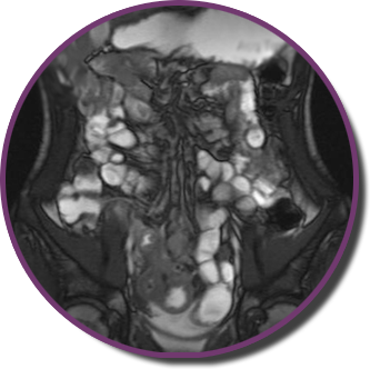
Abdomen MRI
Abdomen MRI with MRCP
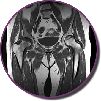
Pelvis MRI
MRI enterography
MRI colonography
© Mag-Medica, All Right Reserved.
Designed By HTML Codex